Anterior Lumbar Interbody Fusion With Robotic-Assisted Percutaneous Screw Placement: A Case Report
By Admin | March 07, 2022
Abstract
Recently, there has been an increase in robotic-assisted spine fusion for degenerative spondylosis of the lumbar spine. We present the case of a 60-year-old female with grade 1 spondylolisthesis at L4/5 and L5/S1 who underwent L4-S1 anterior lumbar interbody fusion (ALIF) with percutaneous robotic-assisted pedicle screw fixation. We provide a detailed analysis of the procedure including the speed of robotic screw placement and pitfalls of this surgical approach.
Introduction
Lumbar spondylolisthesis is an anatomical spinal disorder involving the translation of one vertebral body on another which can cause symptoms of nerve root compression. The management of lumbar spondylolisthesis depends on the etiology, anatomical imaging characteristics, and patient presentation but typically involves lumbar spinal fusion [1-7]. Recently there has been a resurgence in robotic-assisted spinal fusions for a range of spinal disorders resulting in safe and accurate spinal instrumentation [8,9]. We present the case of a 60-year-old female with grade 1 spondylolisthesis where we apply a combination of anterior lumbar interbody fusion (ALIF) with percutaneous robotic-assisted pedicle screw fixation.
Case Presentation
The patient is a 60-year-old female with a past medical history of sarcoidosis and breast cancer treated with surgical resection and chemoradiation that presented with a six-month history of low back pain and dysesthesia radiating down her right posterior thigh and intermittently in the anterior and medial thigh after falling. She denied any left-sided symptoms. She went from ambulating independently to using a walker. She experienced nighttime urinary incontinence due to pain. She failed conservative treatment with lumbar epidural steroid injections, physical therapy, and narcotic pain medications. On examination, she was grade 4+/5 Medical Research Council (MRC) strength in her right hip flexion and knee extension, and 4-/5 right dorsiflexion, plantar flexion, and extensor hallucis longus. She was full strength on the left with normal sensation.
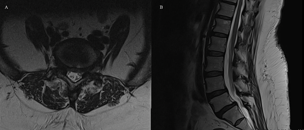
CT of the lumbar spine demonstrated no fractures. MRI of the lumbar spine demonstrated grade 1 spondylolisthesis of L4/5 and L5/S1 with nerve root compression in the neuro-foramen, right greater than left. (Figures 1A, 1B). There was no significant central stenosis. Imaging did demonstrate significant facet arthropathy bilaterally. Upright and flexion-extension x-rays of the lumbar spine re-demonstrated the grade 1 spondylolisthesis, but also showed micro-motion instability at L4/5 and L5/S1. Standing scoliosis x-rays demonstrated minimal levoscoliosis of the lumbar spine. Body habitus precluded spino-pelvic measurements including pelvic incidence, pelvic tilt, and sacral slope.
Given our patient’s unilateral symptomatology, grade 1 spondylolisthesis, and no significant central stenosis, she was offered an L4-5 and L5-S1 ALIF with percutaneous robotic-assisted posterior percutaneous lumbar spinal fusion. A vascular surgeon was used for abdominal access.
Operative procedure
Both the anterior approach and posterior fusion were performed under general anesthesia. The patient was supine for the anterior approach. We utilized a vascular surgeon to gain retroperitoneal access to the anterior spine. A bent spinal needle was...(More)
For more info please read, Anterior Lumbar Interbody Fusion With Robotic-Assisted Percutaneous Screw Placement: A Case Report, by Cureus


